Welcome to our March Newsletter! We continue to innovate with our new lab automation solution, ALH for FlowCam, to increase sample throughput and productivity. Thinking about adding automation to your FlowCam? Check out our white paper on what to consider for successful integration.
And: We're happy to be back to in-person conferences - see our list below for where and when to meet our team to discuss your applications and observe FlowCam in action.
Enjoy reading!
Your FlowCam Team
PRODUCT NEWS
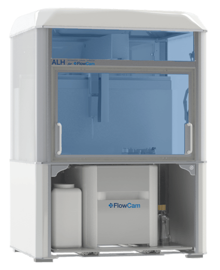
Introducing the New Automated Liquid Handler (ALH) for FlowCam
Designed with flexibility in mind, ALH for FlowCam integrates seamlessly with the FlowCam 8000 instrument series and VisualSpreadsheet software.
Experience unattended FlowCam operation that encompasses time and labor savings around sample preparation, injection, data acquisition, and automated cleaning between samples.
Five Key Benefits of Automating Flow Imaging Microscopy
ALH for FlowCam is designed to help laboratories meet their needs for automation, analysis, and flexibility.
Our new whitepaper describes in detail how automated liquid handling can provide significant productivity and quality improvements that benefit both users and lab operations.
Images of Lindulodinium polyedra shown with red boxes that identify the fluorescence excitation of these dinoflagellates, captured with FlowCam 8400 using a 10X objective
The Ensenada Center for Scientific Research and Higher Education in Baja California was alerted to the early stages of a harmful algal bloom in All Saints Bay. They needed to measure concentration and size distribution and identify the organisms causing the bloom.
After running water samples submitted to our analytical lab, we obtained clear images of the dinoflagellate responsible for the bloom, Lingulodinium polyedra, shown above inside red boxes indicating fluorescence excitation. FlowCam analysis provided the ability to separate different taxa into classifications and individual concentrations.
WHAT WE'RE READING
Preventing Embolism from Medical Implants by Using FlowCam Imaging Particle Analysis
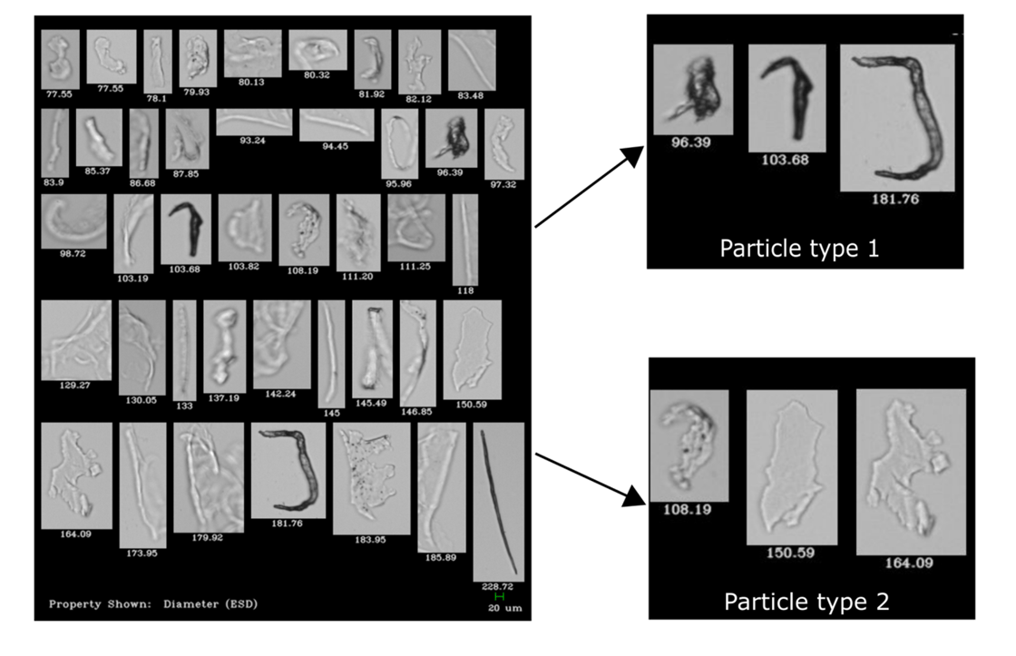
Images captured by FlowCam 8100 reveal fundamental differences in morphological characteristics of particles providing information about their composition and their source.
A team of scientists at Rostock University Medical Center recently used a FlowCam 8100 instrument to analyze the morphology of the subvisible particles shed from a commercially available coronary stent delivery system. The goal of the study was to determine which particle characteristics were most relevant in order to identify the source of the particles.
Using FlowCam images, they were able to draw conclusions about particle origin based primarily on intensity values (degree of transparency), circularity, aspect ratio, and equivalent spherical diameter of the particles as measured by FlowCam.
The Importance of Correct Aperture Settings in Submicron Particle Imaging
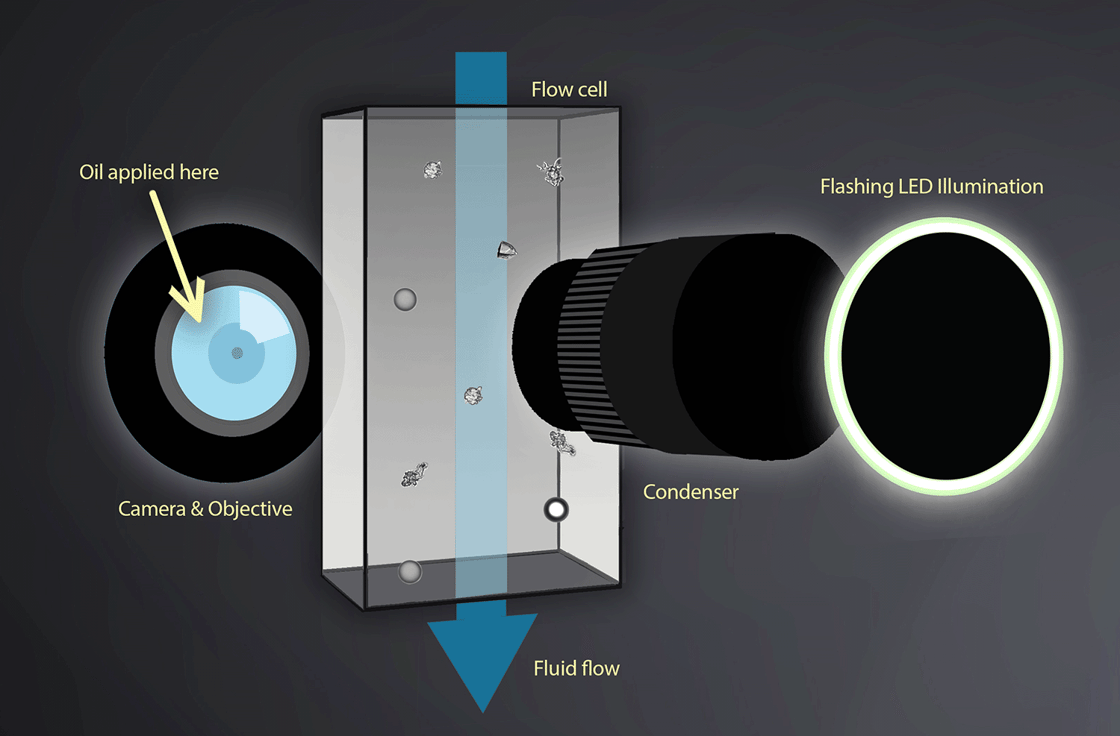
An internal schematic of FlowCam Nano
Numerical Aperture gives the user control over the depth of field and light intensity of the image. When using Flow Imaging Microscopy (FIM) to analyze particles under 2 µm, clear images, accurate sizing, and consistent categorization are crucial, and all depend on the numerical aperture (NA) setting. This setting, along with the wavelength of light, determines how highly resolved the images will be.
In a recent blog post, Chris Mills, one of our engineers, provides a detailed explanation of why this is important to FlowCam Nano.

.png?upscale=true&width=920&upscale=true&name=Lingulodinium%20polyedra%20bioluminescent%20(2).png)